
A dentist checks gum health primarily by inserting a thin, calibrated instrument called a periodontal probe into the space between each tooth and the surrounding gum tissue. This clinical process, formally known as periodontal probing, is the gold standard for detecting gum disease before symptoms appear. The depth of each pocket tells the dentist whether your gum tissue is healthy, inflamed, or actively losing its attachment to the bone beneath. Combined with bleeding assessments and dental X-rays, the exam gives a complete picture of your periodontal condition. Knowing how dentist checks gum health removes the mystery from those numbers your hygienist calls out during your visit.
What are gum pockets and how are they measured?
The gum sulcus is the natural crevice that runs around the base of every tooth where gum tissue meets enamel. In a healthy mouth, this crevice is shallow and firm. When bacteria accumulate and inflammation sets in, the tissue detaches from the tooth and the crevice deepens into what clinicians call a periodontal pocket.
Dentists measure these pockets using a periodontal probe, a slender metal instrument marked in millimeter increments. The probe slides gently below the gumline at six distinct points around each tooth: the mesiobuccal, mid-buccal, distobuccal, mesiolingual, mid-lingual, and distolingual sites. This six-point method captures the full circumference of each tooth's attachment.
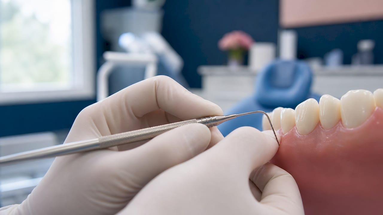
The numbers your hygienist calls out follow a clear clinical scale. Probing depths of 1–3 mm indicate healthy tissue, 4–5 mm signals early disease, and 6 mm or more points to advanced disease requiring prompt intervention. That scale is not arbitrary. It reflects how far bacteria can penetrate and how much tissue has detached from the root surface.
A complete mouth records measurements at all six sites per tooth. For a full set of 28 teeth, that produces 168 individual measurements entered into a periodontal chart. That chart becomes the clinical baseline your dentist compares at every future visit to track whether your gum health is stable, improving, or declining.
| Pocket depth | Clinical meaning | Typical action |
|---|---|---|
| 1–3 mm | Healthy sulcus | Routine maintenance |
| 4–5 mm | Early periodontal disease | Closer monitoring, improved home care |
| 6 mm or more | Advanced disease | Deep cleaning or specialist referral |
Pro Tip: Ask your hygienist to read your numbers aloud and explain which teeth are flagged. Patients who understand their own chart are far more consistent with home care between visits.
The numbered sequence your hygienist calls out during the exam represents the six measurements around each tooth, and it simply maps gum attachment rather than signaling an emergency. Knowing that context turns an anxiety-inducing ritual into useful data you can act on.
How does bleeding on probing signal gum inflammation?
Bleeding on probing, abbreviated as BOP, is the body's immediate response to bacterial irritation in the gum tissue. When the probe touches inflamed tissue, blood vessels close to the surface rupture within 30 seconds. Healthy tissue does not bleed because the vessel walls are intact and the tissue is firm.
Clinicians use standardized cutoffs to interpret BOP scores. Less than 10% of bleeding sites indicates a healthy mouth, 10–30% points to localized gingivitis, and more than 30% signals generalized gingivitis requiring active treatment. These thresholds turn a simple yes-or-no observation into a quantified disease marker.
The reliability of BOP as a diagnostic tool is well established. Absence of bleeding indicates healthy gum tissue 96–99% of the time, making it one of the most sensitive negative predictors in periodontal diagnosis. That means a non-bleeding site is a strong sign of tissue health, not just a lucky result.
A common patient misconception is that bleeding during probing means the hygienist is being too rough. The opposite is true. Bleeding is the tissue telling you it is inflamed. Healthy gums do not bleed when probed with standard force. If your gums bleed consistently, that is clinical evidence of active inflammation, not a sign of a heavy-handed exam.
- BOP occurs within 30 seconds of contact with inflamed tissue
- Scores above 30% indicate generalized gingivitis
- Absence of bleeding predicts health with 96–99% accuracy
- BOP is recorded site-by-site, not as a single whole-mouth score
- Improving your BOP score between visits is a measurable sign that home care is working
Pro Tip: Improving your flossing routine for two weeks before a dental visit does reduce surface inflammation, but it does not eliminate deeper pocket disease. Your dentist will still detect it through probing depth.
What role do X-rays and visual exams play in gum assessment?
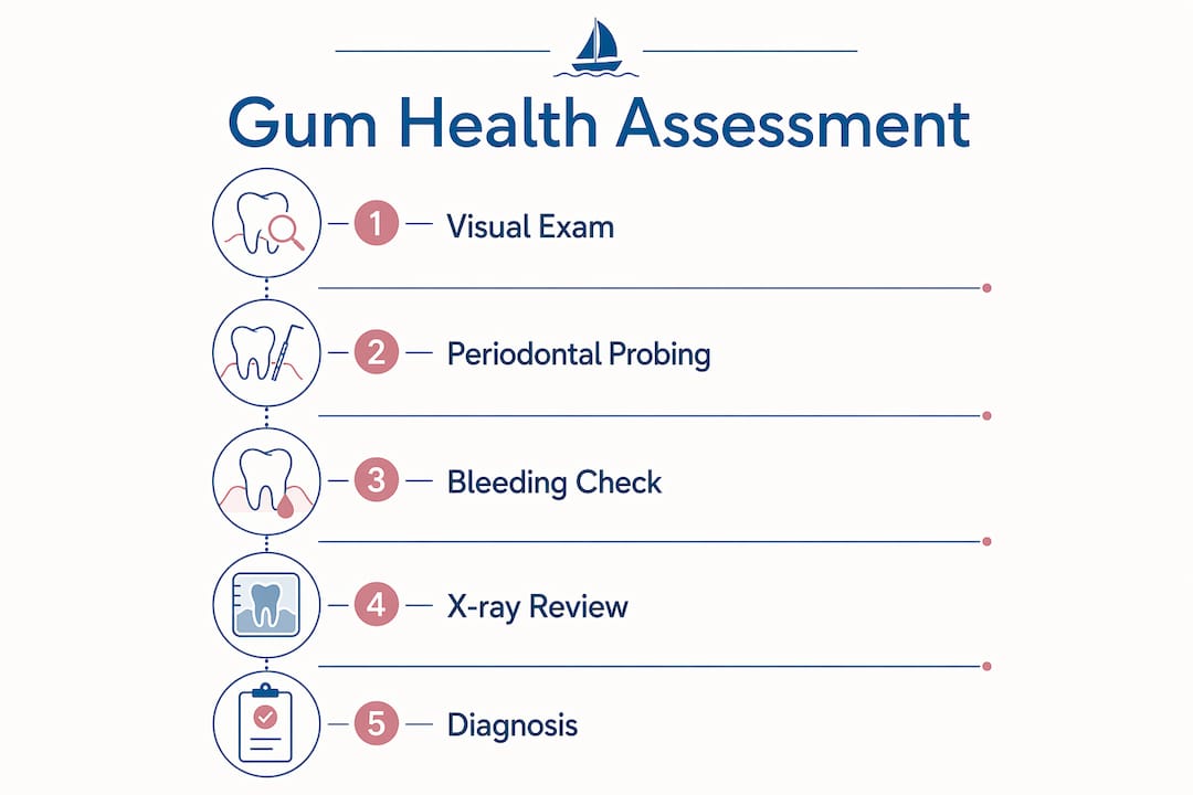
Periodontal diagnosis is never built on probing alone. Dentists combine probing data with a visual clinical exam and dental X-rays to form a complete picture of your gum and bone health. Each tool reveals something the others cannot.
The visual exam comes first. Your dentist inspects gum color, contour, and texture before the probe touches a single tooth. Red, swollen, or receded gum tissue signals inflammation. Visual gum inflammation correlates with deeper pocket findings in 71% of patients, confirming that what the eye sees and what the probe measures tend to agree. That correlation makes the visual exam a reliable first filter.
X-rays add a dimension that neither the eye nor the probe can access directly: the bone supporting each tooth root. Radiographs reveal the height and pattern of bone loss around each tooth. Horizontal bone loss on X-rays suggests generalized disease, while vertical bone loss points to aggressive, localized disease. That distinction directly shapes the treatment plan.
X-rays do have a critical limitation. Bone loss only appears on radiographs after 30–40% of the mineral content has already been destroyed. That means X-rays miss early-stage disease. Probing catches it first. This is why probing remains the primary early detection method, with X-rays serving as confirmation and staging tools.
The full diagnostic picture combines all three inputs:
- Visual exam: gum color, swelling, recession, and bleeding patterns
- Periodontal probing: pocket depths and attachment levels at 168 sites
- Dental X-rays: bone height, bone loss pattern, and root anatomy
Gum disease diagnosis relies on synthesizing data from all these sources rather than any single measurement. The result is a disease stage classification that guides whether you need monitoring, a deep cleaning, or a referral to a periodontist.
How often should you have a gum health assessment?
The American Academy of Periodontology recommends that all patients receive a comprehensive periodontal examination at least once per year, regardless of whether symptoms are present. That recommendation exists because early gum disease produces no pain and no visible warning signs that patients can detect on their own.
A routine dental checkup is not the same as a full periodontal assessment. Standard checkups use rapid screening tools while a formal periodontal assessment provides detailed, tooth-by-tooth mapping of every attachment site. Many patients leave routine visits believing their gums were thoroughly evaluated when only a basic screen was performed.
Early gum disease is reversible, but once bone loss occurs, it is permanent. A full periodontal exam acts as an early warning system that catches disease at the stage when it can still be reversed with professional cleaning and improved home care. Waiting for pain or visible swelling means waiting until the damage is already done.
Regular assessments also produce a personalized record of your gum health over time. Your dentist uses that record to guide home care recommendations, adjust cleaning intervals, and detect any sites that are worsening before they require invasive treatment. Knowing your gum health baseline gives both you and your dentist a concrete target to maintain.
- Annual comprehensive periodontal exams are the professional standard
- Routine checkups do not replace full periodontal assessments
- Early detection prevents irreversible bone loss
- Your periodontal chart is a longitudinal health record, not a one-time snapshot
- Patients with risk factors like diabetes or smoking may need more frequent assessments
Pro Tip: When scheduling your next dental visit, specifically ask whether a full periodontal chart will be recorded. If your practice only does a basic screen, request the complete six-point probing exam. You are entitled to that level of detail.
Key Takeaways
A dentist checks gum health through periodontal probing, bleeding assessment, and X-rays combined, and no single measurement alone determines your diagnosis.
| Point | Details |
|---|---|
| Probing depth scale | Depths of 1–3 mm are healthy; 4–5 mm signals early disease; 6 mm or more requires treatment. |
| 168-site full chart | A complete mouth exam records six measurements per tooth, creating a detailed baseline for tracking change. |
| Bleeding on probing | BOP above 30% indicates generalized gingivitis; absence of bleeding predicts health 96–99% of the time. |
| X-ray limitations | Radiographs detect bone loss only after 30–40% mineral destruction, so probing catches disease earlier. |
| Annual exam standard | The American Academy of Periodontology recommends a full periodontal exam at least once per year. |
What I've learned from watching patients react to their probing numbers
Most patients sit through the probing exam in silence, nodding along while numbers are called out, and then leave with no idea what any of it meant. That gap between clinical data and patient understanding is one of the most preventable problems in dentistry.
The number that surprises people most is not a 6 or a 7. It is a 4. Patients assume that anything under a 5 is fine. It is not. A 4 mm pocket is a clinical flag. It means the tissue has already started to detach, and without intervention, it will deepen. Knowing that one fact changes how seriously patients take their home care.
The other thing I have noticed is that patients who ask questions during the exam leave with better outcomes. When you ask "which teeth are flagged?" or "is my BOP score better than last time?", you turn a passive procedure into a conversation. Your dentist and hygienist want that engagement. It makes their job easier and your gum health better.
The probing exam is not something done to you. It is information collected for you. The numbers belong to you. Understanding them is the first step toward preventing gum disease before it costs you bone, teeth, or both.
— Alex
Gum health assessments at Starboarddental
Starboarddental in Kennebunk offers thorough periodontal assessments that go beyond a basic checkup. The clinical team records full six-point probing charts, evaluates bleeding patterns, and reviews radiographs to build a complete picture of your gum health. From there, care plans are tailored to your specific findings, whether that means a routine cleaning, a referral for scaling and root planing, or a closer monitoring schedule.

Starboarddental's approach combines clinical precision with a genuinely comfortable environment, including ceiling-mounted TVs and same-day appointments for patients who need prompt attention. If you have not had a full periodontal exam recently, or if you are unsure whether your last visit included one, the general dentistry team at Starboarddental is ready to give your gums the thorough evaluation they deserve.
FAQ
What does a dentist use to check gum health?
A dentist uses a periodontal probe, a calibrated metal instrument, to measure the depth of the space between each tooth and the surrounding gum tissue at six sites per tooth.
Are bleeding gums during probing a sign of disease?
Yes. Bleeding on probing is a direct indicator of gum inflammation caused by bacterial buildup. Healthy gum tissue does not bleed when probed with standard clinical force.
How deep should gum pockets be?
Healthy gum pockets measure 1–3 mm. Depths of 4–5 mm indicate early periodontal disease, and 6 mm or more signals advanced disease that requires active treatment.
Can I detect gum disease at home?
No. Early gum disease produces no pain and causes no visible changes that patients can reliably detect. A professional periodontal exam is the only way to identify disease before it causes irreversible bone loss.
How is a full periodontal exam different from a routine checkup?
A routine checkup uses a basic screening approach, while a full periodontal exam records six probing measurements per tooth across all 168 sites, producing a detailed clinical map of your gum attachment levels.