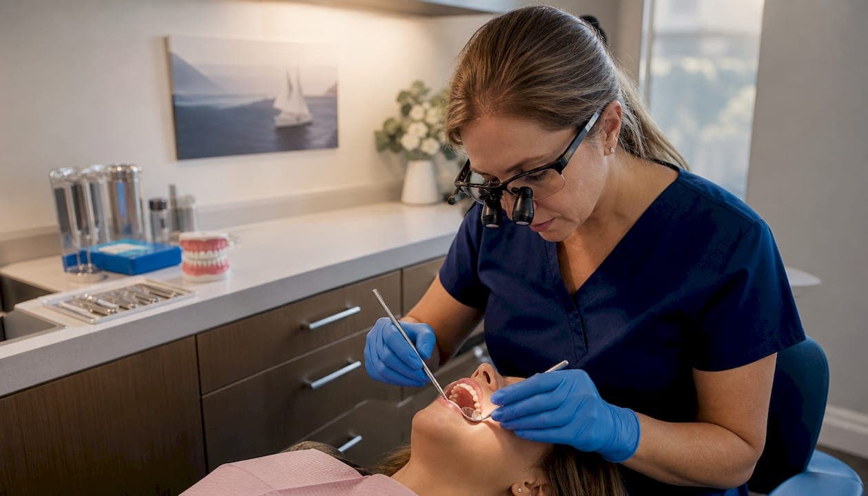
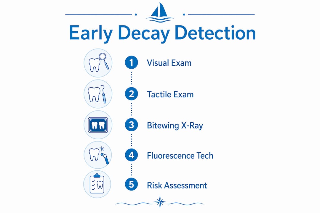
Early tooth decay, known clinically as dental caries in its initial stages, is defined as the gradual demineralization of tooth enamel caused by acid-producing bacteria. Understanding how dentists spot early decay gives you a real advantage: treatment at this stage is faster, cheaper, and far less invasive than treating a full cavity. Dentists use a combination of visual inspection, tactile probing, and radiographic imaging to catch decay before it becomes a problem. Combining all three methods raises diagnostic accuracy to 85–95%, compared to just 50–60% for visual examination alone. That gap explains why a thorough professional exam does what a bathroom mirror never can.
How dentists spot early decay during a visual exam
The first visible sign of tooth decay is a white spot lesion. This is a chalky, opaque patch on the enamel surface caused by mineral loss. It does not hurt, it does not look like a hole, and the most patients walk past it for years without knowing it is there.
Dentists identify these spots through careful visual inspection, but the technique matters. Drying the tooth surface before looking is a critical step. Wet enamel masks the contrast between healthy gloss and the dull, chalky appearance of a demineralized spot. A dry tooth reveals what moisture hides.
Not every discolored spot signals decay. Dentists distinguish between harmless staining from coffee or tea and the structural changes of early caries. Stains tend to follow surface grooves and have a brown or yellow tone. White spot lesions appear as flat, opaque patches, often near the gumline or between teeth.
- White spot lesions: Chalky, opaque patches indicating enamel demineralization
- Surface staining: Brown or yellow discoloration without structural change
- Pitting or roughness: Textural changes felt during tactile probing
- Gumline lesions: Common near the margin where plaque accumulates
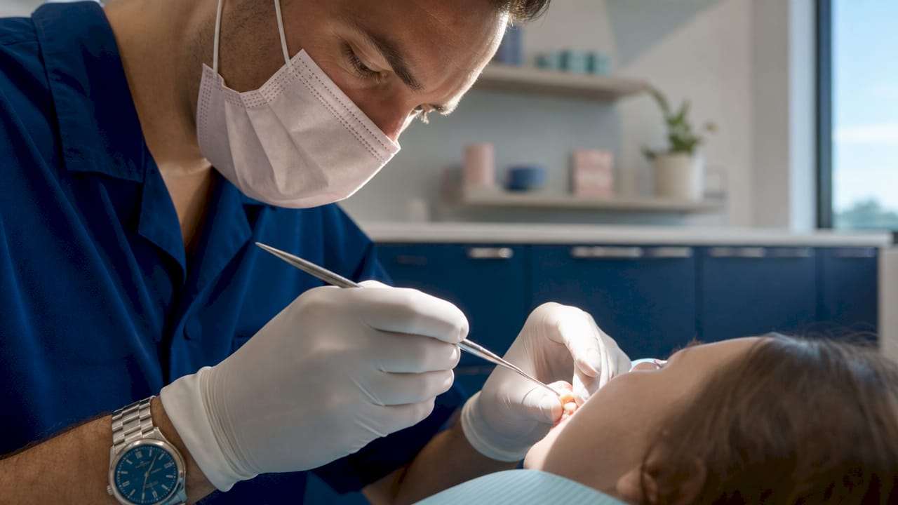
Tactile examination adds a second layer of information. Dentists use a dental explorer, a thin metal probe, to gently feel the tooth surface. Healthy enamel feels smooth and firm. Early decay feels slightly soft or sticky, a sign that the mineral structure has weakened.
Pro Tip: You can notice new white spots at home by looking at your teeth in bright light after brushing. Any chalky patch that was not there before is worth mentioning at your next appointment.
How do dentists use X-rays and imaging to find hidden decay?
Visual and tactile exams have one significant blind spot: the spaces between teeth. Decay that starts in these contact areas, called interproximal decay, is invisible from the outside until it has already progressed. Bitewing X-rays are the standard tool for finding it.
A bitewing X-ray shows the crowns of upper and lower teeth on the same image. Mineral loss appears as a dark shadow within the tooth structure. Dentists read these shadows to assess how deep the decay has traveled and whether it has reached the dentin layer beneath the enamel.
X-rays do have a limitation worth knowing. They cannot detect decay until a meaningful amount of mineral has already been lost. Very early lesions, still at the white spot stage, may not show up on a standard X-ray at all. That is where advanced imaging tools fill the gap.
| Imaging method | What it detects | Limitation |
|---|---|---|
| Bitewing X-ray | Interproximal and deep decay | Misses very early mineral loss |
| Intraoral camera | Surface cracks and visible lesions | Cannot see beneath the surface |
| QLF / DIAGNOdent | Microscopic mineral loss, early caries | Not available in all practices |
Fluorescence technologies like QLF and DIAGNOdent detect mineral loss at the microscopic level, before any shadow appears on an X-ray. These devices shine a specific wavelength of light onto the tooth. Healthy enamel fluoresces differently than demineralized tissue, and the device reads that difference. They are particularly useful for high-risk patients and for monitoring lesions that are too early to treat but too suspicious to ignore.
Pro Tip: Ask your dentist which imaging tools they use and how often they recommend X-rays for your specific risk level. Patients with a history of frequent cavities often benefit from annual bitewing X-rays rather than the standard two-year interval.
Why early detection changes your treatment options
The difference between catching decay at the white spot stage and catching it as a full cavity is the difference between a fluoride treatment and a drill. Early decay is often painless, which is exactly why patients delay seeking care. By the time a tooth hurts, the decay has usually reached the dentin or pulp, and the treatment required is far more involved.
When a dentist identifies a white spot lesion, the first response is not always to fill it. Modern dentistry favors remineralization over drilling wherever possible. Fluoride treatments, prescription-strength toothpaste, and dietary changes can reverse early mineral loss and restore enamel strength without any drilling at all.
The decision pathway dentists follow looks roughly like this:
- Identify the lesion stage. Is it a white spot, or has it broken through the enamel surface?
- Assess the patient's risk level. High sugar intake, dry mouth, and poor hygiene all accelerate progression.
- Choose a treatment approach. Early lesions get a "watch and remineralize" plan. Lesions that have broken through enamel require a dental filling.
- Schedule monitoring. The dentist sets a follow-up interval based on how quickly the lesion appears to be changing.
- Reassess at each visit. If remineralization is working, the lesion may stabilize or reverse. If it progresses, treatment escalates.
Delayed detection removes options. A cavity that reaches the pulp requires a root canal. A tooth that fractures around deep decay may need a crown or extraction. The conditions treated at that point are far more complex than a simple white spot ever was.
What can patients do to support early decay spotting?
Self-assessment has real limits. Patients can observe new white spots and sensitivity at home, but these observations cannot replace a professional diagnosis. What you notice at home is a signal, not a verdict.
That said, knowing what to watch for makes you a better partner in your own care. Early signs worth reporting to your dentist include:
- New chalky or white patches on any tooth surface
- Sensitivity to cold, sweet foods, or air that was not there before
- Food consistently getting caught between the same two teeth
- A rough or sticky feeling on a tooth that used to feel smooth
- Any visible darkening or pitting in a groove or fissure
Good daily habits slow decay before it starts. Brushing twice daily with fluoride toothpaste, flossing once a day, and limiting sugary or acidic foods between meals all reduce the acid exposure that drives enamel breakdown. Staying hydrated supports saliva production, which is the body's natural defense against acid. You can find more specific guidance on preventing cavities and gum disease to build a routine that fits your life.
Pro Tip: Before your dental appointment, make a short list of any new sensitivities or spots you have noticed. Dentists can focus their exam on areas you flag, which improves the chance of catching something early.
How do dentists track early decay over time?
Accurate detection starts before the dentist even looks at your teeth. Thorough cleaning and drying of each tooth surface before examination is a non-negotiable step. Plaque and debris obscure early lesions. A polished, dry tooth surface gives the clearest possible picture.
Tracking decay over time requires more than a single snapshot. Dentists use digital records and, in some practices, subtraction radiography to compare X-rays taken at different appointments. Subtraction radiography overlays two images and highlights any change in density, making it possible to see whether a lesion is growing, stable, or reversing. That level of detail turns a routine X-ray into a monitoring tool.
Key factors that shape how often your dentist schedules exams and imaging:
- Cavity history: Patients with frequent past cavities need more frequent monitoring
- Diet and lifestyle: High sugar intake or acid reflux raises decay risk significantly
- Dry mouth: Reduced saliva from medications or health conditions accelerates enamel breakdown
- Oral hygiene quality: Inconsistent brushing and flossing creates conditions where decay progresses faster
- Age and tooth anatomy: Deep grooves in molars trap bacteria and are harder to clean
Risk-based exam scheduling is the standard approach in modern dental practice. A low-risk patient with no history of decay may need X-rays every two to three years. A high-risk patient may need them annually or more often. The goal is to catch any change before it becomes irreversible.
Key Takeaways
Catching tooth decay at the white spot stage gives dentists the option to reverse it without drilling, making early detection the single most valuable step in preventive dental care.
| Point | Details |
|---|---|
| Combined methods are most accurate | Visual, tactile, and imaging exams together reach 85–95% diagnostic accuracy. |
| White spots are the first warning sign | Chalky enamel patches are reversible with fluoride if caught before they break through. |
| X-rays find what eyes cannot | Bitewing X-rays reveal interproximal decay hidden between teeth. |
| Pain is a late symptom | Early decay causes no discomfort, so waiting for pain means waiting too long. |
| Risk-based scheduling matters | Exam and imaging frequency should match your individual decay risk, not a fixed calendar. |
What I've learned from watching patients avoid the drill
After years of following dental health closely, one pattern stands out: the patients who avoid major restorative work are not the ones with perfect teeth. They are the ones who show up consistently and tell their dentist about small changes they have noticed.
The white spot lesion is the most underappreciated finding in dentistry. It looks like nothing. It feels like nothing. But it is the moment when the entire outcome of that tooth is still negotiable. Dentists who take the time to dry each surface, probe carefully, and use fluorescence tools when available are giving their patients a genuine advantage. Patients who dismiss a chalky spot because it does not hurt are giving decay a head start.
The combination of methods matters more than any single tool. A visual exam alone misses too much. X-rays alone miss the earliest lesions. Fluorescence devices alone are not yet standard everywhere. But a dentist who uses all three, and who tracks changes over time with digital records, is practicing at the level where decay rarely wins.
My honest advice: do not wait for sensitivity or pain to schedule an exam. Tooth sensitivity that you have been ignoring is already a late signal. The earlier you go, the more options you keep.
— Alex
Starboarddental's approach to catching decay before it becomes a cavity
Starboarddental in Kennebunk offers general dentistry services built around early detection and conservative care. The practice uses digital imaging and thorough clinical exams to identify decay at its earliest stage, giving patients more treatment options and fewer invasive procedures.

The team at Starboarddental tailors exam frequency and imaging to each patient's actual risk level, not a one-size-fits-all schedule. Same-day appointments mean you do not have to wait weeks when you notice something new. Whether you are due for a routine checkup or have a specific concern, Starboarddental's approach puts early detection first, so small problems stay small.
FAQ
What is the earliest sign of tooth decay a dentist can detect?
The earliest detectable sign is a white spot lesion, a chalky patch of demineralized enamel. At this stage, decay is often reversible with fluoride treatment and improved oral hygiene.
How do dentists diagnose tooth decay between teeth?
Dentists use bitewing X-rays to detect interproximal decay, which appears as a dark shadow indicating mineral loss. This type of decay is invisible during a standard visual exam.
Can early tooth decay be reversed without a filling?
Yes. Decay caught at the white spot stage can often be reversed through remineralization using fluoride treatments, prescription toothpaste, and dietary changes, with no drilling required.
How often should I get dental X-rays for early decay detection?
Frequency depends on your individual risk level. Low-risk patients may need X-rays every two to three years, while high-risk patients may need them annually.
Does early tooth decay always cause pain or sensitivity?
Early decay is typically painless. Pain and sensitivity usually appear only after decay has progressed into the dentin or pulp, which is why professional exams are the only reliable way to catch it early.