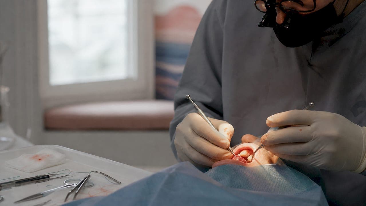
Receding gums cannot grow back on their own, but treatment can stop the progression and, when tissue loss is significant, restore the gumline using grafts or biomaterial alternatives. According to Cleveland Clinic guidance, options range from desensitizing agents and dental bonding for mild cases to surgical grafting for more severe recession. The right path depends on how much tissue you have lost, what caused it, and whether the problem is still active.
Three things to do right now:
- Switch to a soft-bristle toothbrush and ease up on pressure today. Mechanical trauma is one of the most common drivers of recession, and continuing to brush hard will undermine any treatment.
- Book a dental exam. A clinician needs to probe and measure the recession before any treatment plan makes sense. Gum health support starts here.
- Use a desensitizing toothpaste with potassium nitrate or stannous fluoride in the meantime to manage sensitivity from exposed roots.
If you are in the Kennebunk area, Starboarddental offers periodontal evaluations and can walk you through every option from scaling and root planing to restorative bonding.
Key Takeaways
Gum recession cannot reverse itself, but the right treatment sequence, starting with cause correction and scaling, then grafting when needed, can halt progression and restore tissue.
| Point | Details |
|---|---|
| Gums cannot regrow on their own | Treatment halts progression; grafting or biomaterials restore lost tissue when recession is significant. |
| Fix the cause before surgery | Active infection, aggressive brushing, or bruxism must be controlled first or any graft will fail. |
| CTG vs. biomaterial alternatives | A 2026 RCT found comparable 6-month root coverage for CTG and collagen matrices; CTG still leads on long-term data. |
| Nonsurgical care works for mild cases | Scaling, bonding, desensitizers, and brushing correction are sufficient for stable, shallow recession. |
| Starboarddental for local care | Starboarddental in Kennebunk offers periodontal evaluation, scaling, bonding, and treatment coordination for gum recession patients. |
Table of Contents
- What is gum recession and how do you know you have it?
- What causes gum recession?
- How dentists diagnose and classify gum recession
- Nonsurgical gum recession treatment options and when they are enough
- Surgical treatments: which graft or technique is right for you?
- Recovery timeline, outcomes, and what can go wrong
- What does gum recession treatment cost in the U.S.?
- Daily habits that prevent recession from getting worse
- How to choose the right dentist or periodontist for gum treatment
- What the 2026 evidence says about graft alternatives
- What to expect at Starboard Dental for gum recession care
- Starboard Dental is ready to help with your gum health
- Sources
What is gum recession and how do you know you have it?
Gum recession, clinically called gingival recession, is the apical migration of the gingival margin, meaning the gum tissue pulls away from the tooth and exposes the root surface below the enamel-covered crown. That exposed root is softer than enamel and more vulnerable to decay, sensitivity, and further wear.
Symptoms to check for:
- Tooth sensitivity, especially to cold, hot, sweet, or acidic foods and drinks
- Teeth that look longer than they used to
- A visible yellowish or darker band at the base of a tooth (the root surface)
- A notch you can feel with your tongue near the gumline
- Food trapping between teeth more than before
- Bleeding when brushing or flossing, particularly at the gumline
- Tooth mobility in advanced cases where bone loss has also occurred
Mild recession often causes no pain at all, which is why many patients only notice it when a dentist points it out during a routine exam. Sensitivity is usually the first symptom that prompts someone to seek care.
Seek immediate care if you notice: rapid visible change over days or weeks, pus around a tooth, severe or throbbing pain, or a tooth that feels loose. These signs suggest active infection or significant bone involvement that needs prompt attention.
What causes gum recession?
Understanding the cause matters because treatment that does not address the underlying driver will fail or relapse. There is rarely a single cause; most patients have two or three overlapping factors.
Primary causes:
- Periodontal (gum) disease. Bacterial plaque triggers chronic inflammation that destroys the attachment fibers and bone supporting the tooth, pulling the gum margin down with it. This is the most common cause worldwide.
- Aggressive brushing. Scrubbing with a hard-bristle brush or using a horizontal sawing motion abrades the gum margin directly. The result is often a wedge-shaped or V-shaped notch at the gumline, typically on the outer (buccal) surface of premolars and canines.
Predisposing and contributing factors:
- Thin gingival biotype and genetics. Some people simply have thinner, more fragile gum tissue with less underlying bone. This tissue recedes more easily under any mechanical or inflammatory stress.
- Tobacco use. Smoking and smokeless tobacco impair blood flow to gum tissue, suppress the immune response, and accelerate attachment loss.
- Orthodontic tooth position. Moving teeth outside their natural bone envelope can thin the buccal plate and increase recession risk. A comprehensive PMC review explains that orthodontic repositioning can actually prevent or improve recession in specific cases by moving roots into thicker bone, but it can also worsen it when thin buccal bone is present.
- Lip and tongue piercings. Jewelry that repeatedly contacts the gum margin causes localized mechanical trauma and recession, usually on the lingual surface of lower front teeth.
- Bruxism and occlusal trauma. Grinding or clenching generates lateral forces that stress the periodontal attachment and can accelerate recession, particularly when combined with thin tissue.
- Poorly controlled diabetes. Systemic conditions that impair healing and amplify inflammatory responses make periodontal disease more aggressive and recession harder to manage.
How cause shapes treatment: Active infection from periodontal disease must be controlled with scaling and root planing before any graft is placed. A graft placed over active disease will fail. Similarly, if aggressive brushing is the driver, behavior change is non-negotiable before surgery. Grafting over an unchanged brushing habit is a short-term fix at best.
Pro Tip: If your recession is on the outer surface of a single tooth with a notch at the gumline and no recession on adjacent teeth, that pattern strongly suggests brushing trauma rather than periodontal disease. Ask your clinician whether the pattern fits a mechanical cause, because the treatment path is different.
How dentists diagnose and classify gum recession
A proper diagnosis goes beyond a visual glance. Clinicians use several tools to measure severity, identify the cause, and determine whether nonsurgical care or surgery is appropriate. You can read a detailed walkthrough of how a dentist checks gum health before your appointment.
Standard diagnostic steps:
- Medical and dental history. Medications, systemic conditions like diabetes, tobacco use, and orthodontic history all affect both cause and treatment planning.
- Visual examination. The clinician assesses gum color, contour, tissue thickness, and the position of the gumline relative to the cementoenamel junction (CEJ).
- Periodontal probing. A calibrated probe measures recession depth in millimeters from the CEJ to the gum margin, plus pocket depth and attachment loss at six points per tooth.
- Clinical photography. Baseline photos document the extent of recession and are used to track treatment outcomes over time.
- Radiographs. X-rays are taken when bone loss is suspected, since bone support determines whether grafting alone is sufficient or whether more complex periodontal treatment is needed first.
Classification systems: The Miller classification (Classes I through IV) has been used for decades to predict how much root coverage is achievable with grafting. Class I and II recessions, where there is no bone or soft-tissue loss in the interdental space, typically allow complete root coverage. Class III allows only partial coverage. Class IV, with severe bone and tissue loss, offers limited coverage potential. More recent classification systems, such as the Cairo RT1/RT2/RT3 descriptors, are increasingly used in academic and specialist settings and map similarly to treatment predictability.
What determines surgical versus nonsurgical care: Clinicians look at recession depth and width, the width of remaining attached (keratinized) gingiva, tissue thickness, whether the recession is progressing, and the patient's aesthetic concerns. Shallow, stable recession with adequate attached gingiva and no sensitivity often needs only behavior modification and monitoring. Progressive recession, thin or absent keratinized tissue, root sensitivity that does not respond to desensitizers, or aesthetic concerns typically indicate a referral to a periodontist for grafting.
Nonsurgical gum recession treatment options and when they are enough
Nonsurgical care is the right starting point for most patients. It addresses the cause, manages symptoms, and in mild cases may be all that is needed.
Core self-care measures:
- Use a soft-bristle manual or electric toothbrush. Medium and hard bristles are not necessary for plaque removal and cause direct tissue damage.
- Brush at a 45-degree angle to the gumline with short, gentle strokes. The Merck Manual specifically recommends this technique alongside desensitizing toothpaste for managing recession.
- Floss daily using a gentle C-shape technique. Snapping floss into the gum causes trauma.
- Stop tobacco use. Smoking cessation is one of the highest-impact changes a patient can make for periodontal health.
- Wear a night guard if you grind or clench. Bruxism-related occlusal forces accelerate recession and will undermine any treatment.
Clinical nonsurgical options:
- Scaling and root planing (deep cleaning). When periodontal disease is driving recession, scaling and root planing removes calculus and bacterial deposits from below the gumline and smooths root surfaces to reduce reattachment of bacteria. This is the foundation of nonsurgical periodontal therapy.
- Topical antimicrobials and antibiotics. Locally delivered agents like chlorhexidine chips or minocycline microspheres can be placed in periodontal pockets after scaling to reduce bacterial load in targeted sites.
- Desensitizing agents and fluoride varnish. Professionally applied fluoride varnish and desensitizing agents block exposed dentinal tubules and reduce sensitivity. These do not restore tissue but provide meaningful symptom relief.
- Dental bonding. Tooth-colored composite resin can be applied to cover an exposed root surface, reducing sensitivity and improving appearance. Dental bonding is a reversible, relatively low-cost option for patients who are not surgical candidates or who want a cosmetic improvement without grafting.
- Orthodontics. When tooth malposition is contributing to recession, moving the tooth back into the bone envelope can reduce recession risk and, in some cases, improve the gumline. This is rarely a standalone fix for established recession but can be part of a combined plan.
When nonsurgical care is usually sufficient: Mild, stable recession (1–2 mm) with adequate attached gingiva, no active infection, and manageable sensitivity often responds well to behavior modification, scaling, and desensitizers alone. The key word is stable. If the recession is not progressing and the patient is not bothered by sensitivity or appearance, monitoring with regular cleanings is a reasonable approach.
Red flags that mean a periodontist referral:
- Recession depth of 3 mm or more
- Absent or minimal keratinized gingiva
- Progressive recession despite behavior change
- Sensitivity that does not respond to desensitizing toothpaste after 4–6 weeks
- Aesthetic concerns the patient wants addressed
Pro Tip: Before agreeing to surgery, ask your dentist: "Can we try desensitizing agents, bonding, and a brushing technique correction for 6–8 weeks and then re-evaluate?" For mild recession, that sequence often resolves sensitivity and buys time to confirm whether the recession is truly progressing.
Simple treatment flow:
- Correct the cause (brushing technique, tobacco, bruxism management, plaque control).
- Complete scaling and root planing if periodontal disease is present.
- Apply desensitizers or bonding for symptom relief.
- Re-evaluate at 6–8 weeks. If recession is stable and symptoms are controlled, continue monitoring.
- If recession is progressing, tissue is thin, or aesthetics are a concern, refer to a periodontist for surgical evaluation.
Surgical treatments: which graft or technique is right for you?
When nonsurgical care is not enough, surgery can restore lost gum tissue and cover exposed roots. The options have expanded significantly, and the choice between them depends on how much tissue needs to be replaced, where it is, and how much donor-site discomfort the patient is willing to accept.
Connective tissue graft (CTG)
The connective tissue graft remains the most predictable surgical option for root coverage. The surgeon takes a small piece of connective tissue from beneath the palatal mucosa (the roof of the mouth) and places it under a flap at the recession site. The palate heals over, and the graft integrates at the recipient site.
CTG is the benchmark against which other techniques are measured. It works well for Miller Class I and II recessions and can achieve complete root coverage in favorable cases. The tradeoff is donor-site morbidity: the palate is sore for 1–2 weeks, and some patients find this the hardest part of recovery.
Free gingival graft (FGG)
The free gingival graft takes a strip of tissue directly from the palatal surface (including epithelium) rather than from beneath it. It is used primarily to increase the width of attached keratinized gingiva rather than for aesthetic root coverage, because the color match is less predictable. It is a good choice when the goal is tissue augmentation around a tooth with very little attached gingiva.
Coronally advanced flap (CAF) and pedicle flap
These techniques reposition existing gum tissue rather than harvesting new tissue. The coronally advanced flap slides the patient's own gum margin up to cover the recession. It works best when there is enough tissue to move and is often combined with a CTG or biomaterial for better outcomes. A pedicle flap rotates adjacent tissue laterally to cover a recession site.
Tunneling technique
The tunnel approach passes the graft material under the gum tissue through small incisions rather than creating a large open flap. It preserves blood supply to the tissue, which can improve graft integration, and tends to produce good aesthetic results. It is technically demanding and not appropriate for every anatomy.
Biomaterial alternatives: AlloDerm and Geistlich Mucograft
Two widely used alternatives to palatal tissue harvesting are AlloDerm (an acellular dermal matrix, or ADM, derived from processed human donor skin) and Geistlich Mucograft (a porcine-derived collagen matrix). Both eliminate the need for a palatal donor site, which is the primary source of postoperative discomfort in CTG procedures.
A 2026 randomized controlled trial found comparable short-term root coverage between CTG and collagen matrices for localized type 1 recession at six months. That is encouraging for patients who want to avoid palate harvesting. The caveat is that six months is a short follow-up window. CTG has decades of long-term data behind it; collagen matrices and AlloDerm have a shorter evidence track record for sustained outcomes.
| Technique | Donor-site morbidity | Root coverage predictability | Recovery | Best indications |
|---|---|---|---|---|
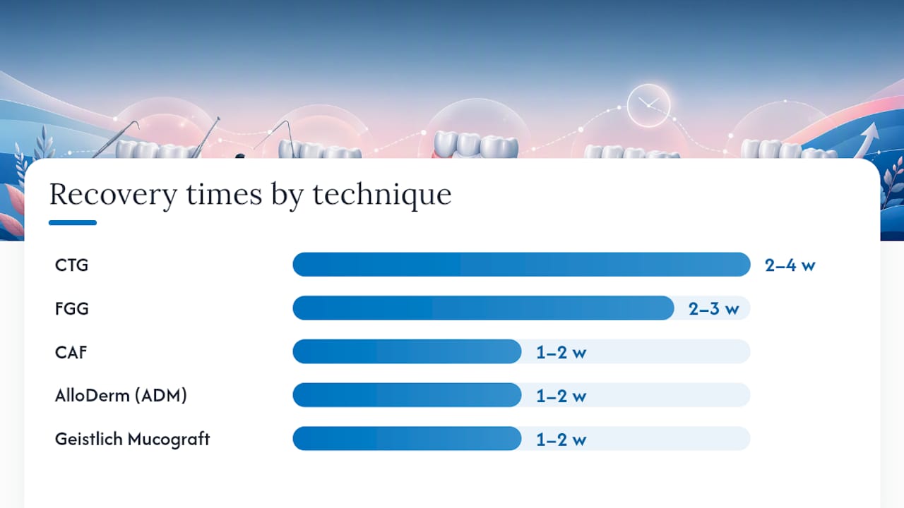
| Connective tissue graft (CTG) | Moderate (palate) | High; gold standard | 2–4 weeks | Miller/RT1–RT2; single or multiple teeth |
| Free gingival graft (FGG) | Moderate (palate) | Moderate for coverage; high for tissue augmentation | 2–3 weeks | Thin tissue; keratinized tissue augmentation |
| Coronally advanced flap (CAF) | None (no harvest) | Good when combined with CTG or matrix | 1–2 weeks | Adequate existing tissue; often combined |
| AlloDerm (acellular dermal matrix) | None | Comparable short-term; less long-term data | 1–2 weeks | Patients avoiding palate harvest; multiple sites |
| Geistlich Mucograft (collagen matrix) | None | Comparable at 6 months per 2026 RCT | 1–2 weeks | Localized recession; patients avoiding palate harvest |
| Tunneling technique | Depends on graft used | Good aesthetic outcomes | 1–2 weeks | Favorable anatomy; aesthetic zone |
When recession is associated with non-carious cervical lesions (NCCLs), the notched root surface that results from abrasion, a 2026 systematic review and meta-analysis found that combined periodontal-restorative treatment reduced dentin sensitivity faster than periodontal treatment alone, though long-term differences between the two approaches were small. Restoring the notch with composite before or alongside grafting prevents ongoing abrasion and complements the graft.
Pro Tip: At your surgical consultation, ask three specific questions: What percentage of root coverage do you expect in my case? Will you be harvesting from my palate, and if so, how do you manage donor-site pain? Can you show me clinical photos of similar cases you have treated? A surgeon who cannot answer these questions clearly is not the right fit.
Recovery timeline, outcomes, and what can go wrong
Recovery from gum grafting is manageable for most patients, but it requires discipline, especially in the first two weeks.
Typical recovery milestones:
- 24–72 hours: Swelling peaks, especially if a palatal donor site was used. Ice packs applied externally for 20 minutes on and 20 minutes off help. Soft foods only. Avoid rinsing vigorously.
- Days 3–7: Discomfort at the donor site (if CTG) is usually the most uncomfortable period. Over-the-counter pain relievers manage it for most patients. The recipient site typically causes less pain.
- Week 2: Sutures are often removed or dissolve. The graft site looks pink and slightly raised. Gentle brushing resumes around the graft with a soft brush.
- 1–3 months: The graft integrates and the tissue matures. Final aesthetic result is not fully visible until 3–6 months post-surgery.
Factors that influence success: Smoking is the single biggest modifiable risk factor for graft failure. Poorly controlled diabetes impairs healing and increases infection risk. Oral hygiene during recovery matters enormously. Patients who follow postoperative instructions closely, stay off tobacco, and attend follow-up appointments have substantially better outcomes.
Signs of complications that need prompt attention:
- Graft appears white or gray and is not reattaching (graft failure)
- Increasing rather than decreasing pain after day 3
- Pus or foul taste around the surgical site
- Persistent bleeding beyond 24 hours
- Fever
Statistic callout: A 2026 randomized controlled trial reported comparable short-term root coverage improvements between connective tissue grafts and collagen matrices at six months for localized type 1 recession, suggesting biomaterial alternatives are a viable option for patients who want to avoid palate harvesting.
For patients managing sensitivity from exposed roots during recovery, tips for sensitive teeth can help bridge the gap while the graft matures.
What does gum recession treatment cost in the U.S.?
Costs vary widely depending on the number of teeth involved, the procedure type, and the practice's location and fee schedule. These are general ballpark figures, not guarantees.
Practical steps to estimate your out-of-pocket cost:
- Ask the practice for an itemized treatment estimate with CDT codes before committing.
- Call your insurer with those codes and ask what your plan pays, what your deductible is, and whether you have hit your annual maximum.
- Request a pre-authorization (also called a predetermination) in writing before scheduling surgery. This is not a guarantee of payment but gives you a written estimate of what the insurer will cover.
- If your recession is linked to a systemic condition like diabetes, ask whether any portion of treatment might be submitted under your medical insurance rather than dental.
- Ask about phased treatment if cost is a barrier. Treating the most severe or symptomatic sites first spreads cost across benefit years.
Daily habits that prevent recession from getting worse
Prevention is far easier than repair. Whether you have had treatment or are trying to avoid it, these habits protect the gum margin.
Do:
- Brush with a soft-bristle brush at a 45-degree angle to the gumline, using short circular or back-and-forth strokes with light pressure. An electric toothbrush with a pressure sensor removes the guesswork.
- Use a desensitizing toothpaste with potassium nitrate or stannous fluoride. Both ingredients reduce sensitivity from exposed roots; stannous fluoride also has antimicrobial properties.
- Floss daily using a gentle technique. Proper flossing removes plaque from the gumline where brushing cannot reach.
- Schedule professional cleanings every 3–6 months if you have a history of periodontal disease. Standard twice-yearly cleanings may not be frequent enough.
- Wear a night guard if you grind or clench. Bruxism generates forces that stress the gum attachment and accelerate recession.
Avoid:
- Hard-bristle or medium-bristle brushes.
- Brushing immediately after acidic foods or drinks (wait 30 minutes).
- Tobacco in any form.
- Lip or tongue piercings that contact the gum margin.
- Ignoring a new or growing sensitivity. Sensitivity is often the first warning sign.
Orthodontic treatment can reduce recession risk when tooth malposition is a contributing factor. Moving roots into thicker bone reduces the mechanical stress on thin gingival tissue. This works best as a preventive measure or early intervention; established recession usually still requires periodontal grafting even after orthodontic correction.
How to choose the right dentist or periodontist for gum treatment
For mild recession, a general dentist can manage scaling, bonding, and behavior counseling. For progressive recession, absent keratinized tissue, or surgical grafting, a periodontist is the right specialist.
When to see each provider:
- General dentist: Mild, stable recession; scaling and root planing; bonding; night guard fabrication; monitoring and referral.
- Periodontist: Progressive recession; surgical grafting (CTG, FGG, tunneling, biomaterials); complex multi-tooth cases; patients with active periodontal disease needing specialist-level management.
- Oral surgeon: Occasionally involved in complex bone grafting cases, but most soft-tissue recession surgery is handled by periodontists.
Questions to ask at a consultation:
- Are you a board-certified periodontist, and how many gum graft procedures do you perform per year?
- What percentage of root coverage do you expect in my specific case, and what classification is my recession?
- Will you be using my own tissue, AlloDerm, or a collagen matrix like Geistlich Mucograft, and why?
- Can I see before-and-after photos of similar cases?
- What is your complication rate for this procedure?
- What does the follow-up schedule look like, and what happens if the graft does not fully take?
- What is the full cost estimate, and does your office handle pre-authorization?
Look for a practice with digital imaging for precise diagnosis, clear communication about options, and a willingness to discuss both surgical and nonsurgical paths. Sedation options matter for anxious patients. Starboarddental's periodontics services cover the full range from evaluation through treatment coordination.
What the 2026 evidence says about graft alternatives
The question patients and clinicians are asking most often right now: can a collagen matrix or allograft replace connective tissue from the palate without sacrificing long-term results?
The short answer is: for short-term outcomes, probably yes. A 2026 randomized controlled trial found comparable root coverage improvements between CTG and collagen matrices for localized type 1 recession at six months. That is a meaningful finding because it suggests patients who want to avoid palate harvesting have a credible alternative for localized cases.
The longer answer requires more caution. Six months is not long enough to draw conclusions about durability. CTG has decades of follow-up data showing stable outcomes.
The practical tradeoff comes down to patient priorities. If avoiding donor-site pain is the priority and the recession is localized and early-stage, a collagen matrix or AlloDerm is a defensible choice with current evidence. If maximum long-term predictability is the priority, especially for multiple teeth or deeper recession, CTG remains the benchmark.
Useful sources for further reading:
- PubMed 41495722: The 2026 RCT comparing CTG and collagen matrices at six months.
- PMC4944726: A comprehensive review of recession causes, types, and the role of orthodontic treatment.
- Springer/BMC Oral Health 2026 meta-analysis: Systematic review of combined periodontal-restorative therapy for recession with NCCLs.
What to expect at Starboard Dental for gum recession care
At Starboarddental, the approach to gum recession starts with a thorough exam, not a rushed recommendation. Every patient gets periodontal probing, clinical photos, and a clear explanation of what the measurements mean before any treatment is discussed. The goal is to understand the cause first, because a plan that does not address the driver will not hold.
For most patients, that means starting conservatively: correcting brushing technique, completing any necessary scaling and root planing, and monitoring the response. When surgical options are indicated, the team coordinates with specialist referrals and integrates restorative care, such as bonding or crown work, when recession is associated with root notching or cosmetic concerns.
The clinic uses digital scanners for precision, and the environment is designed to reduce anxiety, with ceiling-mounted TVs, personalized comfort items, and a team that takes the time to explain each step. Same-day appointments are available for patients with acute symptoms.
Note: Formal clinical credentials and any local case documentation will appear in the published version of this article.
Starboard Dental is ready to help with your gum health
Gum recession is one of those conditions where waiting makes the treatment more complex and more expensive. Starboarddental offers the full range of care that recession patients need: periodontal evaluations, scaling and root planing, dental bonding for exposed roots, night guards for bruxism, and coordination with restorative care when NCCLs are involved.

New patients in Kennebunk, York, and Saco, ME can book a comprehensive exam and get a clear, itemized picture of what treatment would look like for their specific case. Bring your insurance card, a list of any medications, and any previous dental records if you have them. The first appointment covers diagnosis, cause identification, and a phased treatment plan so you know exactly what comes next and what it will cost.
Schedule your gum health exam at Starboarddental and get a straight answer about where your gums stand and what to do about it.
Sources
The following sources were used to build this article and are worth consulting directly for more detail:
- Randomized controlled trial: CTG versus collagen matrices (PubMed 41495722)
- Gingival recession: its causes and types, and the importance of orthodontic treatment - PMC
- Gum Recession: Causes, Prevention, Surgery & Treatment
- Gum recession - Merck Manual Consumer Version
- Efficacy of combined periodontal-restorative treatment versus periodontal treatment alone for gingival recession associated with non-carious cervical lesions: a systematic review and meta-analysis
This article is general information, not a substitute for advice from a qualified doctor. Consult a qualified healthcare professional about your own circumstances before acting on anything here.