Dental X-rays are safe for the vast majority of patients because the radiation dose involved is small and the images are ordered only after a clinical exam justifies them. The main exceptions worth knowing: cone beam CT (CBCT) delivers a meaningfully higher dose than a standard bitewing, unnecessary repeat imaging should always be avoided, and children need stricter justification before any scan. A single intraoral X-ray exposes you to roughly what you'd absorb from a small amount of ordinary background radiation, which is why dentists follow the ALARA and ALADA principles: as low as reasonably achievable, and as low as diagnostically acceptable.
TL;DR:
- Most dental X-rays involve minimal radiation, with intraoral images delivering only about 1 to 8 microsieverts, comparable to a few hours of natural background exposure.
- Cone beam CT scans can produce significantly higher doses, potentially equaling several days or weeks of background radiation, so their use should be carefully justified and limited to specific diagnostic needs.
- Children and pregnant women require stricter justification and smaller fields of view due to higher radiation sensitivity, and routine imaging should be avoided unless clinically necessary.
- Modern dental practices follow ALARA and ALADA principles by using digital sensors, limiting scan volume, and avoiding unnecessary repeat images to keep exposure low.
- Responsible imaging involves a clinical exam review, prior record consultation, and patient-specific dose minimization strategies before any X-ray is performed.
Table of Contents
- Dental X-Ray Safety: Understanding the Different Imaging Types
- How Much Radiation Do Dental X-Rays Actually Deliver?
- Who Needs Extra Caution: Children, Pregnancy, and Frequent Imaging
- How Dental Offices Keep Radiation Exposure Low
- When CBCT Makes Sense (and When It's Overkill)
- What Radiation-Conscious Dental Care Looks Like in Practice
- Schedule Digital X-Rays With a Practice That Explains Every Scan
- Sources
- FAQ
Dental X-Ray Safety: Understanding the Different Imaging Types
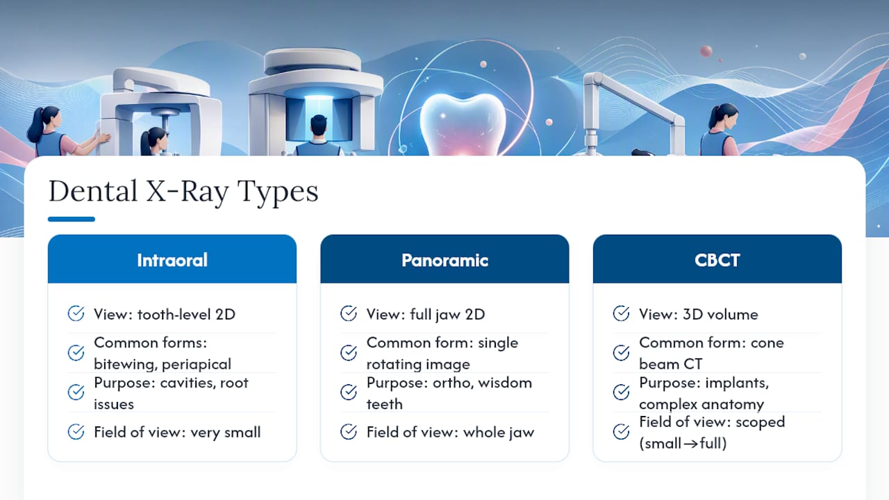
Not all dental X-rays are the same, and the type your dentist orders says a lot about both the diagnostic question and the radiation involved. Intraoral X-rays, the small films or sensors placed inside your mouth, come in two common forms: bitewings, which capture the crowns of upper and lower teeth together to catch cavities between teeth, and periapical images, which show a full tooth from crown to root tip to check for infections, abscesses, or bone loss around the root.
Panoramic X-rays work differently. A single rotating image captures your entire jaw, all your teeth, your sinuses, and the temporomandibular joints in one pass. Dentists use these for orthodontic planning, wisdom tooth evaluation, and getting a wide-angle view before more detailed treatment.
Then there's cone beam CT, or CBCT, which produces a three-dimensional image rather than a flat 2D picture. It's the imaging of choice for dental implant planning, complex root canal anatomy, evaluating bone volume, and diagnosing issues that a flat image simply can't resolve, like the exact position of an impacted tooth relative to a nerve canal.
The field of view matters as much as the modality itself. A small, focused CBCT scan of a single tooth delivers far less radiation than a full-jaw scan, and a dentist who understands this will scope the image to answer only the question at hand. None of this happens in a vacuum, either. Responsible imaging follows a clinical exam and a review of your prior radiographs, not a routine calendar reminder. If your dentist already has a periapical image from eight months ago showing the area in question, that record should factor into whether a new one is even needed.
How Much Radiation Do Dental X-Rays Actually Deliver?
The numbers are smaller than most patients assume, and they vary by exam type in ways worth knowing before your next appointment. A single intraoral radiograph, whether bitewing or periapical, delivers an effective dose of roughly 1 to 8 microsieverts (μSv). A panoramic exam runs higher, typically 4 to 30 μSv, because it captures a much larger area in one exposure. Cephalometric X-rays, the side-profile images orthodontists use for jaw and bite planning, sit in a narrow band around 2 to 3 μSv.
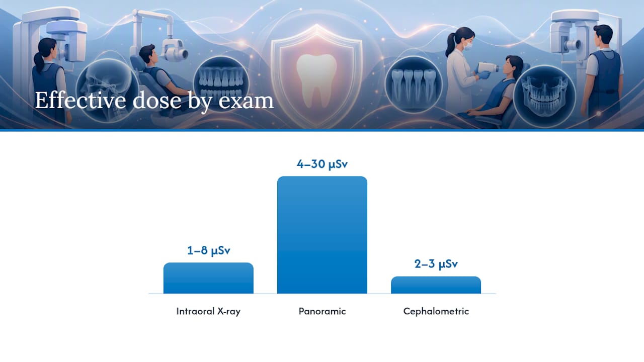
CBCT is the outlier. Depending on the field of view and scan protocol, effective doses can vary widely, from moderate to substantially higher doses than other dental X-rays, according to a CBCT dose review. That spread is wide because CBCT dose depends heavily on scan volume, not just on the fact that it's 3D imaging.
Context helps make these numbers meaningful. Average daily exposure to natural background radiation, from soil, cosmic rays, and even the potassium in your own body, runs in the range documented by the Nuclear Regulatory Commission. A single intraoral X-ray falls well under a full day's worth of that ambient exposure. A panoramic exam is closer to a couple of days' worth. CBCT can equal several days to a couple of weeks of background exposure, depending on the scan size, which is precisely why dentists don't reach for it casually.
| Exam type | Typical effective dose | Rough background radiation equivalent |
|---|---|---|
| Bitewing / periapical (intraoral) | 1–8 μSv | Hours of background exposure |
| Panoramic | 4–30 μSv | About 1–3 days of background exposure |
| Cephalometric | 2–3 μSv | A few hours of background exposure |
| CBCT (small to large field of view) | Tens to hundreds of μSv | Several days to a couple of weeks |
A few factors drive the variation within each category:
- Equipment age and calibration: newer digital systems require less radiation to produce a diagnostic image than older analog units.
- Exposure settings (kVp, mA, exposure time): even small adjustments change the dose delivered per image.
- Field of view: a targeted CBCT scan of one quadrant uses far less radiation than a full-jaw volume.
- Patient anatomy: technique sometimes needs adjusting for jaw size or tissue density, which affects exposure calculations.
Cumulative exposure over a lifetime of dental visits stays low precisely because each individual exam sits in the microsievert range. The IAEA's technical guidance on diagnostic reference levels exists so clinics can benchmark their equipment against known safe ranges and catch outliers before they become a pattern.
Who Needs Extra Caution: Children, Pregnancy, and Frequent Imaging
Radiation sensitivity isn't uniform across every patient, and this is where blanket reassurance stops being useful. Children absorb radiation differently than adults do. Their tissues are still developing, and pediatric sensitivity to ionizing radiation is substantially higher than in adults, according to research on pediatric radiation sensitivity. That's the biological reason pediatric dentists apply stricter selection criteria: fewer routine films, smaller fields of view when CBCT is genuinely needed, and imaging timed to actual clinical findings rather than a fixed recall schedule.
Pregnancy is a different kind of caution; for practical concerns about dental care abroad or during pregnancy, learn more about dental coverage abroad for expats. Dental treatment, including necessary X-rays, is generally considered safe during pregnancy when the imaging is clinically indicated, but the standard advice still holds: tell your dentist you're pregnant before any imaging is scheduled, so the team can weigh necessity and apply appropriate shielding and collimation.
Frequent imaging deserves its own scrutiny. Some situations genuinely call for repeat X-rays at shorter intervals: active periodontal disease being monitored for bone loss, orthodontic treatment tracking root movement, or a root canal being followed to confirm healing. Routine imaging on a fixed annual schedule regardless of your oral health history is a different story, and it's exactly what the ADA's newer guidance pushes back against.
Here's what to do at your next visit if you want to stay proactive about this:
- Bring or request transfer of prior radiographs so a new office isn't duplicating recent images.
- Ask directly why a specific X-ray is being recommended and what it's expected to show.
- Request the smallest field of view that still answers the clinical question, especially for CBCT.
- Mention pregnancy, recent medical imaging, or any relevant health history before imaging is scheduled.
Pro Tip: Keep a simple log of your dental X-ray dates and types, especially if you switch practices often. It takes thirty seconds and gives every new dentist the context they need to avoid ordering a redundant scan.
How Dental Offices Keep Radiation Exposure Low
Two principles govern responsible dental imaging: ALARA (as low as reasonably achievable) and ALADA (as low as diagnostically acceptable). Neither means avoiding X-rays altogether. Both mean an image only gets taken when it will change or confirm a clinical decision, and when it is taken, the dose is optimized to the minimum needed for a readable result.
In practice, that principle translates into specific equipment and protocol choices, not just a philosophy on a wall poster. Here's what a radiation-conscious office actually does:
- Uses digital receptors instead of film. Digital sensors require 80 to 90 percent less radiation than traditional film to produce a usable image, and the file appears instantly, cutting down on retakes from processing errors.
- Applies rectangular collimation. Shaping the X-ray beam to match the size of the receptor, rather than using a wider round beam, reduces the tissue volume exposed without sacrificing image quality.
- Relies on receptor holders. These positioning devices keep the sensor stable and correctly angled, which cuts down on blurry or poorly angled images that would otherwise require a retake.
- Follows technique charts. Standardized exposure settings for different patient sizes and tooth locations, rather than guesswork, keep every exposure at the minimum needed dose.
- Runs routine equipment QA. Calibration checks catch machines drifting toward higher output before that drift becomes a pattern across dozens of patients.
One detail surprises a lot of patients: the ADA's updated guidance notes that lead aprons and thyroid collars are no longer routinely recommended for many dental imaging procedures. That's not a step backward. Modern beam collimation and digital sensors already restrict the radiation field so tightly that a lead apron adds little additional protection, and in some CBCT scans it can actually interfere with image quality by casting an artifact into the field. Beam control does more of the protective work than a physical shield ever did.
Pro Tip: If an office still uses film X-rays or doesn't mention reviewing your prior images before a new scan, it's fair to ask why. A modern, radiation-aware practice should have a ready answer for both.
Documented technique charts and well-maintained receptor holders do more than reduce single-exposure dose. They cut down on retakes caused by positioning errors, which is where a surprising amount of cumulative exposure actually comes from over years of dental visits, according to an FDI policy statement on radiation safety.
When CBCT Makes Sense (and When It's Overkill)
CBCT earns its higher dose when a flat 2D image genuinely can't answer the clinical question. Implant planning is the clearest case: knowing the three-dimensional bone volume and the exact location of the nerve canal below it changes where and how an implant gets placed. Complex root canal anatomy, impacted teeth sitting close to critical structures, and jaw pathology are the other common indications where 3D detail changes the treatment plan.
The dose trade-off is real. Where a bitewing sits at 1 to 8 μSv, a CBCT scan can run into the hundreds of μSv depending on the field of view selected, a gap wide enough that it should never be the default choice for a routine cavity check.
Clinicians limit that exposure several ways: selecting the smallest field of view that captures the area of interest, choosing lower-resolution voxel settings when a coarser image still answers the clinical question, and using half-cycle exposure protocols on machines that support them. Field of view and protocol selection are the two biggest levers, since CBCT dose depends heavily on scan volume rather than the technology itself.
A few things worth confirming before agreeing to a CBCT scan:
- Ask what specific finding the scan is expected to confirm that a 2D image couldn't.
- Confirm the smallest field of view is being used for your situation.
- Ask who will interpret the scan, since specialist radiologic review adds a layer of accuracy to complex findings.
Good practice documents the reasoning, not just the image. A note explaining why 3D imaging was chosen over 2D, and why the specific field of view was selected, is part of the shared decision that should happen before the scan, not after.
What Radiation-Conscious Dental Care Looks Like in Practice
In a radiation-conscious dental practice, digital radiography is the standard, and every image request follows a clinical exam and a review of existing records before a new scan gets ordered. That's the ALARA and ALADA framework in action, not just a phrase on an intake form.
Patients should expect a conversation, not a rubber stamp: why a particular X-ray is being recommended, what it's expected to show, and whether a prior image already answers the question. Comfort matters here too. Sensor placement, positioning, and pacing all affect how tolerable an exam feels, especially for anxious patients or younger ones sitting still for a bitewing.
For more on how imaging fits into a broader diagnostic picture, Starboard Dental's guide to spotting early decay walks through what dentists are actually looking for when they order a scan in the first place.
— Alex
Schedule Digital X-Rays With a Practice That Explains Every Scan
A radiation-aware dental practice orders digital X-rays only after an exam establishes real clinical need, and every scan uses low-dose digital sensors rather than film. That distinction matters if you've ever left an appointment unsure why an image was taken at all. Some dental practices offer same-day appointments allowing you to get a diagnostic answer and a treatment plan in one visit instead of waiting weeks for a follow-up slot.

If you're due for a checkup or have questions about how often you personally need imaging, Starboard Dental's general dentistry page covers exams, cleanings, and oral cancer screenings, all with the same clinical necessity standard applied to radiographs. You can also see the full range of services, including digital X-rays and TMD care, on the Starboard Dental homepage. Booking takes a few minutes online, and any imaging questions you have get answered before a single image is taken, not after.
This article is general information, not a substitute for advice from a qualified doctor. Consult a qualified healthcare professional about your own circumstances before acting on anything here.
Sources
- X-Rays/Radiographs | American Dental Association
- Radiation dose information for dental imaging (PMC)
- Radiation doses in dental radiology - FAQs for health professionals | IAEA
FAQ
Should I decline dental X-rays?
Declining outright isn't usually the right move if your dentist has a clinical reason for the image, since untreated decay or bone loss missed without imaging carries far more risk than the low dose involved. It's reasonable to ask what the X-ray is expected to show and whether a recent prior image already covers it.
Are dental X-rays safe now, given newer digital technology?
Yes. Digital sensors cut radiation exposure by 80 to 90 percent compared to older film systems, and a single intraoral image delivers a dose comparable to a few hours of everyday background radiation.
Is radiation exposure from dental X-rays harmful?
At the low doses involved in routine dental imaging, the absolute risk is very small. A 2018 review found some studies suggesting a possible association between dental X-rays and certain cancers in specific cohorts, but the evidence is mixed, and researchers caution against overinterpreting it given how low individual doses actually are.
Is it really necessary to get X-rays at the dentist?
Necessity depends on your individual oral health, not a fixed schedule. X-rays are justified when they help detect cavities between teeth, bone loss, infections, or planning needs for treatments like implants, and a dentist following ALARA/ALADA principles should be able to explain the specific reason behind each one ordered.
How often should I really need dental X-rays?
There's no universal interval that fits every patient. Frequency depends on your cavity risk, gum disease history, and whether you're in active treatment, which is why Starboard Dental bases imaging decisions on an exam and your dental history rather than a routine calendar date.