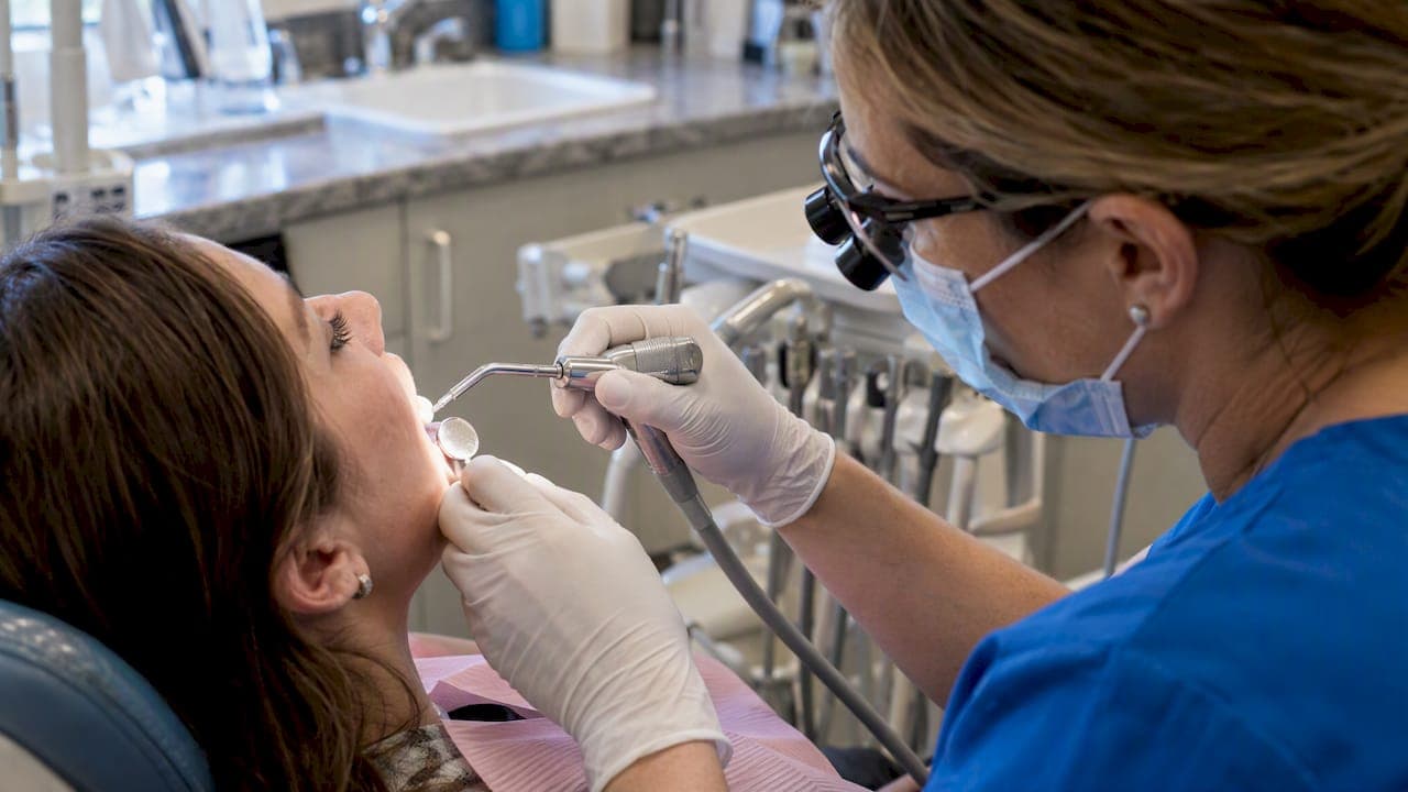
White spots are most often enamel opacities caused by mineral loss or a developmental change in the enamel, and many are either reversible or treatable with conservative care. The CDC links mild fluorosis to excess fluoride during tooth formation, while the ADA's chairside guidance ranks fluoride varnish and resin infiltration among the standard nonrestorative fixes. See a dentist for an activity assessment before deciding on treatment.
TL;DR:
- Active white spots are more likely to become darker or more noticeable when the tooth is dried, indicating ongoing demineralization.
- Fluoride varnish and resin infiltration are the most effective minimally invasive treatments for early lesions, with reapplication often needed over several months.
- Developmental spots from fluorosis or hypoplasia are permanent but can be cosmetically improved with bonding, veneers, or microabrasion.
- Proper diagnosis relies on visual, tactile, and fluorescence exams to determine if a lesion is active, stable, or a developmental defect.
- Preventative measures include daily fluoride use, reducing sugar intake, controlling dry mouth, and thorough cleaning around braces or aligners.
Table of Contents
- What a white spot lesion actually is
- Common causes of white spots on teeth
- How dentists diagnose white spots and check if they're active
- Treatment options for white spots, from least to most invasive
- Preventing white spots and protecting treated enamel
- How Starboard Dental approaches white spot assessment and treatment
- Treat the tooth before you treat the look of it
- Book a white spot evaluation with Starboard Dental
- FAQ
- Sources
What a white spot lesion actually is
A white spot looks chalky or opaque because the enamel underneath has lost some of its mineral content. Healthy enamel is translucent because light passes through its tightly packed crystal structure. When minerals leach out, tiny pores open up inside the enamel, and light scatters off those pores instead of passing through. The tooth looks white or dull in that one area because the refractive index inside the porous enamel no longer matches the surrounding, intact enamel.

Dentists generally sort these into two categories. The first is early tooth decay, often called a white spot lesion, where demineralization from acid and plaque has started eating into the enamel surface but hasn't yet broken through to form a cavity. The second is a developmental defect, like fluorosis or enamel hypoplasia, where the enamel formed with reduced mineral density or thickness before the tooth ever erupted. Both can look nearly identical to the naked eye.
That overlap is why visibility and timing matter during an exam. A lesion that is invisible when the tooth is wet can become obvious the moment a dentist dries it with air, since water fills the enamel's pores and temporarily restores some of its translucency. A dentist weighs the tooth's location, the patient's history, and how the spot behaves under drying before deciding what kind of white spot they're looking at.
Common causes of white spots on teeth
Cause determines the fix, so identifying it is the first real step in treatment.
- Plaque-driven demineralization: Sugars and acids from food feed bacteria that produce acid, and that acid strips minerals from enamel, especially around the gumline or between teeth where brushing misses.
- Orthodontic appliances: Braces create more surfaces for plaque to hide around, and reviews of orthodontic patients report white spot lesions appearing at notably higher rates once brackets are in place, usually clustered near the bracket edges.
- Fluorosis: Too much fluoride during the years permanent teeth are forming, a window that generally closes by age 8 to 10, can leave faint white or paper-white patches once those teeth erupt, according to the CDC.
- Enamel hypoplasia and developmental defects: Illness, nutritional problems, or trauma to a baby tooth during a child's early years can disrupt enamel formation in the permanent tooth growing underneath.
- Dry mouth and mouth breathing: Reduced saliva flow removes one of the mouth's natural buffers against acid, which can accelerate demineralization in people who breathe through their mouth at night or take medications that reduce saliva.
A spot tied to plaque and diet usually responds to remineralization. A spot tied to fluorosis or a developmental defect is permanent in its origin, even though its appearance can still be treated cosmetically.
How dentists diagnose white spots and check if they're active
An exam for a white spot is less about naming the spot and more about figuring out whether it's still progressing. That distinction, active versus inactive, drives everything that follows.
- Visual and tactile exam: The dentist looks at the spot wet, then dries it with air, since a lesion that becomes more visible on drying is typically still demineralizing.
- ICDAS scoring: Many practices use the International Caries Detection and Assessment System to grade the lesion's size and depth on a standard scale, which helps track whether it's getting better or worse over time.
- Fluorescence tools: Devices like laser fluorescence readers can flag subsurface changes that aren't obvious by eye alone, though they're used alongside, not instead of, a visual exam.
- Radiographs: X-rays come into play when a dentist suspects the demineralization has progressed deeper than the enamel surface. Our guide on how dentists spot early decay walks through this in more detail.
- Risk signals: A lesion with a soft, chalky texture, nearby plaque buildup, or cavitation on an adjacent tooth surface points toward active decay rather than an old, stable defect.
An active lesion gets arrested first. A stable, developmental spot can usually move straight to cosmetic options without that intermediate step.
Treatment options for white spots, from least to most invasive
Treatment follows a logical order: stop any active decay, then address what's left cosmetically.
Noninvasive care comes first. For active, noncavitated lesions, professional fluoride varnish is the standard starting point. The ADA's chairside guidance lists 5% sodium fluoride varnish and 1.23% APF gel among the recommended options, often reapplied every few months depending on risk. CPP-ACP pastes and consistent at-home fluoride toothpaste use support the same goal: rebuilding mineral content before the lesion worsens. Our piece on fluoride varnish for adults covers how that schedule typically works outside the pediatric setting.
Resin infiltration bridges the gap between prevention and drilling. The procedure etches the enamel surface, dries it, then infiltrates a low-viscosity resin into the porous lesion body, where it hardens and changes how light passes through the spot. A systematic review and meta-analysis found the resin penetrates a substantial portion of the lesion on average and reliably improves the tooth's appearance, though researchers note long-term color stability still needs more study. A clinical case report on treating fluorosis this way showed clear aesthetic improvement, but also noted the treated area needed extra polishing after staining from turmeric in the patient's diet, a reminder that foods to avoid after teeth whitening for lasting results can impact treatment durability.
Microabrasion removes a very thin layer of surface enamel mechanically, which works for shallow, superficial opacities but isn't suited to deeper lesions since it can't address anything below the surface it removes.
Restorative and cosmetic options come in when the first three don't fully resolve the spot or when a tooth needs more substantial correction. Composite bonding can mask a persistent spot directly, while porcelain veneers cover the whole visible surface for more extensive cases. Tooth contouring sometimes complements either option; see our guide on tooth contouring for what that procedure involves.
- Noninvasive fluoride programs typically run several months of reapplication rather than a single visit.
- Resin infiltration is usually completed in one appointment per tooth.
- Bonding or veneers may take one to two visits depending on the number of teeth involved.
Pro Tip: Ask your dentist to dry-test the spot during your exam. It's the simplest way to see whether a lesion is still active before committing to a treatment plan.
Preventing white spots and protecting treated enamel
Prevention matters whether you're trying to stop a new spot from forming or protect one that's already been treated.
- Brush twice daily with a fluoride toothpaste and clean between teeth daily, since plaque left along the gumline is where most active lesions start.
- Limit how often you snack on sugary or acidic foods and drinks. Frequency matters more than quantity, since each exposure restarts the acid cycle.
- Keep up with professional fluoride varnish at the intervals your dentist recommends, and ask about sealants for children at higher risk of decay.
- If you wear braces or clear aligners, pay extra attention to cleaning around brackets or trays, since both appliances create spots plaque can hide.
- Address dry mouth, whether from medication or mouth breathing, since reduced saliva removes a natural defense against demineralization.
How Starboard Dental approaches white spot assessment and treatment
At Starboard Dental, a white spot evaluation starts with a visual and tactile exam, photo documentation to track the lesion over time, and a risk assessment based on diet, hygiene, and any orthodontic history. Active lesions get a remineralization plan first, typically fluoride varnish paired with hygiene coaching. Once a lesion is stable, patients who want the spot's appearance addressed can move into composite bonding, veneers, or teeth whitening, all available through Starboard Dental's general and cosmetic dentistry services. Appointments are available to help get that first exam scheduled without a long wait.
Treat the tooth before you treat the look of it
The order matters more than most patients expect. An active lesion that gets masked cosmetically before it's arrested will likely keep progressing underneath the fix, which defeats the purpose. Resin infiltration is a genuinely useful tool, but it works best as part of an ongoing prevention routine, not a one-time erase button. Whitening and bonding can both interact with existing white spots in ways that surprise people, sometimes making the contrast more visible rather than less, so it's worth setting realistic expectations before any cosmetic step.
— Alex
Book a white spot evaluation with Starboard Dental
If you've noticed a white spot and aren't sure whether it's decay, fluorosis, or something else, an exam is the fastest way to find out and get a plan that fits your situation.

The practice offers appointments paired with digital scanners that give a precise read on the tooth's surface. Bring photos of the spot if you've tracked how it's changed, since that history helps with the activity assessment. From there, your dentist can map out next steps, whether that's a fluoride program through general dentistry or a cosmetic fix through cosmetic dentistry once any active decay is under control. For spots that call for more extensive repair, restorative dentistry covers that ground too. Call or book online to get the first exam on the calendar.
This article is general information, not a substitute for advice from a qualified doctor. Consult a qualified healthcare professional about your own circumstances before acting on anything here.

FAQ
How do you get rid of white dots on your teeth?
Treatment depends on whether the dot is active decay or a developmental spot like fluorosis. Active lesions usually respond to professional fluoride varnish and better hygiene, while stable spots can often be masked with resin infiltration or covered with bonding or veneers if infiltration alone doesn't fully blend the area.
What do dehydrated teeth look like?
A dried tooth surface often shows white spots more clearly than a wet one, since water normally fills tiny pores in demineralized enamel and restores some translucency. Dentists use this drying effect deliberately during exams to judge whether a lesion is active.
Do white calcium spots on teeth go away?
Spots from mineral loss can improve or disappear with consistent fluoride treatment if caught early, since the enamel can partly remineralize. Developmental spots, like those from fluorosis, do not fade on their own but can be made far less visible with resin infiltration, bonding, or similar cosmetic treatment.
Is it possible to remove white spots?
Yes, though "remove" usually means masking or blending rather than erasing the enamel change itself. Resin infiltration, microabrasion, bonding, and veneers each address this differently depending on how deep and how visible the spot is.
Sources
- CDC — About dental fluorosis
- White spot lesions: diagnosis and treatment – a systematic review
- Systematic review and meta-analysis on infiltrative resins and WSLs
- ADA — Chairside guide: nonrestorative treatments for carious lesions on permanent teeth